Late Diastolic Depolarization
 Late Diastolic Depolarization is characterized by the exponential section of the AP curve. This acceleration in the rate of
change of the curve is due to the activity of the sodium/calcium exchangers (NCX) that are activated by calcium ions. Cytoplasmic calcium
ions are moved out of the cell in exchange for three sodium ions ... a net gain of positive charge.
Late Diastolic Depolarization is characterized by the exponential section of the AP curve. This acceleration in the rate of
change of the curve is due to the activity of the sodium/calcium exchangers (NCX) that are activated by calcium ions. Cytoplasmic calcium
ions are moved out of the cell in exchange for three sodium ions ... a net gain of positive charge.
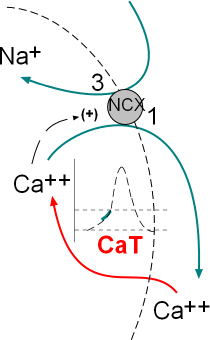
The illustration at the left shows the calcium current, through transient calcium channels (CaT),
and the exchange of three sodium ions for one calcium by sodium/calcium exchangers (NCX). These activities
occur simultaneously; the green section of the AP graph shows the effect on the membrane potential.
Structure of Membrane Channel and Exchanger
Transient Calcium Channels, CaT
These channels have a structure similar to CaL channels described in the Depolarization tutorial
but do not have the beta subunit. Since they are not too abundant the current through them is small. They activate at a relatively
low voltage (~-55mV) and are often referred to as low-voltage activated channels.
Sodium/Calcium Exchanger, NCX
Exchangers are membrane-embedded proteins that move one type of ion through the membrane in exchange for another type of ion. They
are not voltage-dependent but are controlled by the binding of a 'ligand'. A ligand is a substance (usually a small molecule) that forms a
complex with another molecule to serve a biological purpose. In this case the 'ligand' is Ca2+s that bind at the inner-membrane surface
of the exchanger.
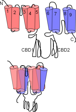
The NCX exchanger has nine membrane-embedded helices connected
by loops of various sizes. The top illustration to the left shows this extended structure. Each helix is called a transmembrane
sequence (TMS); TMS 1-5 constitute 'alpha unit 1' (pink) while TMS 6-9 constitute alpha unit 2 (lavender). Between TMS 2 & 3 and
between TMS 7 & 8 there are short reentrant loops.
A pair of large cytoplasmic loops, CBD1 and CBD2, extend into the cytoplasm; these have Ca2+ binding sites.
The lower illustration shows how the helices are arranged within the membrane; the two
alpha units lying atop one another so that the two reentrant loops are brought close together. Close inspection shows these
two 'hairpins' lined up; Na+ and Ca2+ binding sites are within this region
These exchangers can operate in either a forward (calcium out/sodium in) or a reverse (sodium out/calcium in) mode. The concentrations
of sodium and calcium ions plus the membrane potential itself determine which mode operates.
Late Diastolic Depolarization Currents
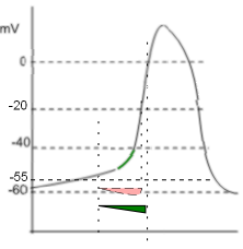
The illustration to the right shows both 'current icons'.
The pink 'icon' represents a calcium current entering the cell through CaT channels. The green 'icon' represents a sodium
current entering due to the activity of sodium/calcium exchangers.
Calcium Current, ICa
The shape of the icon (pink) indicates that these channels begin opening at ~-55mV (first vertical line) and inactivate rapidly
at ~-20mV (second vertical line). Compared to the CaL channels that drive the depolarization of the AP (that open at ~-40mV, 'threshold')
these are low-voltage activated ... -55 is lower than -40. The positive charge brought into the cell via these channels is not the main contributor
to the exponential shape of late diastolic depolarization as described below.
Sodium Current, INa, Forward Mode
The activity of the exchangers depends on several factors. First, there must be a sufficiently high concentration of
cytoplasmic Ca2+ to bind to the CDC1 and CDC2 sites on the exchangers' cytoplasmic loops. The greater the number
of Ca2+ bound to these sites the greater the rate of exchange of 1 Ca2+ for 3 Na+. Each exchanger
can remove as many as 5,000 Ca2+ per second. Accordingly, it is not the build up of Ca2+ from CaT channels
that accounts for the exponential increase in the AP but rather the Na+ that the exchangers bring into the cell.
Additionally, the membrane potential must be negative for the exchangers operate in the 'forward mode' ... calcium ions are moved
out of the cell and sodium ions moved in. The green 'current icon' indicates that the sodium current abruptly stops when the
membrane potential reaches 0mV (third vertical line).
Updated: 1/30/2016
Continue to Calcium Clock
Return to previous tutorial... Early Diastolic Depolarization
Return to home page