Early Diastolic Depolarization
 The heart muscle itself is triggered to beat (systole) due to the accumulation of positive charges at the peak of the pacemaker AP.
The rest period between successive beats is called diastole; during this time the pacemaker membrane potential is undergoing
diastolic depolarization.
The heart muscle itself is triggered to beat (systole) due to the accumulation of positive charges at the peak of the pacemaker AP.
The rest period between successive beats is called diastole; during this time the pacemaker membrane potential is undergoing
diastolic depolarization.
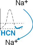
The section of the diagram to the left shows the influx of sodium ions through the HCN channel.
The change in pacemaker membrane potential during this time is a slow, gradual upward slope due to
the influx of sodium ions through hyperpolarization-activated cyclic nucleotide-gated channels (HCN). These channels were originally
called 'funny' channels because they activate during repolarization while other channels activate during depolarization. The influxing sodium current
competes with the effluxing potassium current and is responsible for the 'turnaround' in the AP at the end of repolarization.
Hyperpolarization-activated Cyclic Nucleotide-gated Channels (HCN)
The 'hyperpolarization-activated' part of the channels' name indicates that they, unlike other channels, activate during repolarization.
These channels are additionally unique in that they do not have inactivation gates. They are mixed channels that preferentially allow
the influx of sodium ions but also allow some efflux of potassium ions. They are often called 'pacemaker channels' because the
autonomic nervous system can modify their gating behavior thus changing the heart rate.
Configuration
The mechanisms involved in the gating behavior of these channels is still under investigation. The following is one
of several theories concerning gating and is presented here as a conceptual model.
This channel is composed of four separate proteins arranged in a circle around a central pore. The S1-S4 membrane-embedded
helices (sensor domain) are represented as green cylinders and the pore domain is represented by the yellow cylinders. The S4
helices each have nine positive charges instead of the customary four or five. There is a small helix (light green) on the cytoplasmic S4S5 linker.
The external S5S6 linker has a small helix (yellow) extends down into the pore forming the selectivity filter that determines which
ions may pass. Only two of the four proteins are illustrated here.
Deactivated
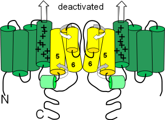 The term 'deactivated' means the activation gates are closed. (Not having an inactivation gate these channels can not 'inactivate'.)
The first illustration shows the deactivated
configuration that exists when the charge at the inner-membrane surface (bottom of illustration) is not sufficiently
negative to hold the S4 sensor helices down (see arrows pointing toward outer-membrane surface).
The small helix (light green) on the S4S5 linker lies horizontally. The S5 and S6 helices are twisted
(curved arrows) in such a way that the cytoplasmic ends of the S6 helices block the pore.
The term 'deactivated' means the activation gates are closed. (Not having an inactivation gate these channels can not 'inactivate'.)
The first illustration shows the deactivated
configuration that exists when the charge at the inner-membrane surface (bottom of illustration) is not sufficiently
negative to hold the S4 sensor helices down (see arrows pointing toward outer-membrane surface).
The small helix (light green) on the S4S5 linker lies horizontally. The S5 and S6 helices are twisted
(curved arrows) in such a way that the cytoplasmic ends of the S6 helices block the pore.
Activated
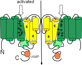 The second illustration shows the activated configuration. The arrows show the S4 helices held down when
the charge at the inner-membrane surface is sufficiently negative to do this. As a result, the cytoplasmic ends of the S6 helices move
and no longer block the pore. One hypothesis suggests that the downward position of the S4 helices push down on one end of the small S4S5 linker
helices (light green) so that their other ends are lifted. This may cause the S5 and S6 helices to twist (curved arrow) and unblock the pore.
The second illustration shows the activated configuration. The arrows show the S4 helices held down when
the charge at the inner-membrane surface is sufficiently negative to do this. As a result, the cytoplasmic ends of the S6 helices move
and no longer block the pore. One hypothesis suggests that the downward position of the S4 helices push down on one end of the small S4S5 linker
helices (light green) so that their other ends are lifted. This may cause the S5 and S6 helices to twist (curved arrow) and unblock the pore.
The orange circles represent binding sites for cyclic AMP (cAMP). When bound, this
cyclic nucleotide ... the 'CN' part of the channels' name ... causes channel activation at a more negative membrane potential thus starting
diastolic depolarization sooner.
Funny (Sodium) Current, If
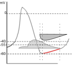 This graph emphasizes the early diastolic depolarization section (red) of the AP curve.
Two 'current icons' are shown indicating that the effluxing potassium current (lighter grey) and the influxing sodium current (darker grey)
occur simultaneously. Keep in mind that the relative position of the horizontal border of a 'current icon' indicates whether the current
is influxing or effluxing. If the current is effluxing, like potassium, the horizontal border is at the bottom; if influxing, like sodium,
it is at the top.
This graph emphasizes the early diastolic depolarization section (red) of the AP curve.
Two 'current icons' are shown indicating that the effluxing potassium current (lighter grey) and the influxing sodium current (darker grey)
occur simultaneously. Keep in mind that the relative position of the horizontal border of a 'current icon' indicates whether the current
is influxing or effluxing. If the current is effluxing, like potassium, the horizontal border is at the bottom; if influxing, like sodium,
it is at the top.
The HCN channels begin activating at ~-55mV (first vertical dashed line) near the end of repolarization.
The shape of the left border of the sodium 'current icon' indicates the maximum current is reached very quickly pushing the membrane potential to
~-60mV (second dashed line). This 'low point' of the AP is called the mean diastolic potential (MDP).
The shape of the right
border of the icon indicates a slowly decreasing current that continues after the potassium current has ended (third dashed line).
In the region between the second and third vertical dotted lines both the potassium and sodium currents are decreasing.
However, because the decrease in the potassium current is faster than the decrease in the sodium current, there is a net gain in
positive charge as indicated by the red region of the AP curve.
After the third dotted line,
when the potassium current has ended, the AP curve begins changing from linear to exponential (curved); this marks the change from early
to late diastolic depolarization.
Updated:2/19/2016
Continue to Late Diastolic Depolarization
Return to previous tutorial... Repolarization
Return to home page