Depolarization
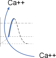 The section of the diagram to the left shows the influx of calcium ions through CaL channels.
This influx is short lived because these ions
bind to calmodulin (Ca/CaM) that increases the rate of channel closing. However, the calcium current can be
extended by a sustained, rapid heart rate that results in channel phosphorylation by calcium/calmodulin-dependent kinase II (CaMKII).
The section of the diagram to the left shows the influx of calcium ions through CaL channels.
This influx is short lived because these ions
bind to calmodulin (Ca/CaM) that increases the rate of channel closing. However, the calcium current can be
extended by a sustained, rapid heart rate that results in channel phosphorylation by calcium/calmodulin-dependent kinase II (CaMKII).
L-type Calcium Channel, CaL
Structure
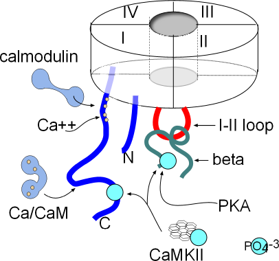 The basic structure of the membrane-embedded portion of the channel has been described in a previous tutorial about
channel structure. In calcium channels, the four six-alpha-helix proteins are linked together forming one long
protein. When linked in this fashion, each of the original four proteins is called a domain (I-IV); they form
a circle around a central pore as shown to the right.
The basic structure of the membrane-embedded portion of the channel has been described in a previous tutorial about
channel structure. In calcium channels, the four six-alpha-helix proteins are linked together forming one long
protein. When linked in this fashion, each of the original four proteins is called a domain (I-IV); they form
a circle around a central pore as shown to the right.
Both ends of this four-domain protein are suspended in the cytoplasm. Both are shown as blue lines in the
illustration; the N-terminal (N) is from domain I and the C-terminal (C) is from domain IV. Also shown in
the cytoplasm is a red loop from domain I to II that forms a 'hinged lid' type of inactivation gate.
Inactivation is voltage-dependent but there are other mechanisms that affect the rate of inactivation.
There are a number of sites where various cytoplasmic components can attach and modify the behavior of this channel.
The C-terminal has two sites where calcium/calmodulin (Ca/CaM) can attach.
CaMKII (calcium/calmodulin-dependent kinase II) can add a phosphate group
(PO4-3) (light blue) at a site near
the proximal end of the C-terminal and also to the beta protein. Phosphokinase A (PKA) can also attach a phosphate group
to the beta protein.
Calcium Current, ICa
The symbol 'I' stands for current ... the net directional flow of charge. In the upcoming tutorials you will see action potential
graphs with a grey 'current icon' superimposed over specific portions of the action potential (AP). In this situation it is positioned
over the depolarization portion (red) of the AP. The width of the icon indicates how long the current lasts and is represented by its horizontal
border. If the current is due to an influx of ions the horizontal border will be the top of the icon; if the current
is effluxing the horizontal line will be the bottom of the icon. This icon shows in influxing current.
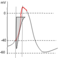 Voltage-dependent activation occurs when the membrane potential reaches ~-40mV (threshold) there is a sudden and strong influx of calcium ions that causes the
upswing (red, depolarization) of the action potential. This point is marked by a vertical line that crosses the threshold
and the beginning of the icon's horizontal border. The shape of the icon indicates there is an instantaneous and large influxing
current ... shape of the left border of icon ... that immediately begins to decline rapidly, slowing slightly just before the current stops...
shape of the right border.
Voltage-dependent activation occurs when the membrane potential reaches ~-40mV (threshold) there is a sudden and strong influx of calcium ions that causes the
upswing (red, depolarization) of the action potential. This point is marked by a vertical line that crosses the threshold
and the beginning of the icon's horizontal border. The shape of the icon indicates there is an instantaneous and large influxing
current ... shape of the left border of icon ... that immediately begins to decline rapidly, slowing slightly just before the current stops...
shape of the right border.
The extensive length of the left border reflects the force of the 10,000:1 calcium concentration gradient that drives this strong current.
The vertical nature of the border indicates that the activation gates of essentially all the channels open fully and simultaneously.
Voltage-dependent inactivation begins at the bottom point of the current icon. The shape of the right border of the icon indicates that
some of the channels' inactivation gates (I-II loops) close immediately, followed by closure of the remaining channels.
When the right border reaches the top horizontal border the current has ended; all channels have closed.
A second vertical line passes through the peak of the action potential and marks the end of depolarization. However, the red
portion of the AP curve indicates that a small calcium current is occurring during the beginning of repolarization.
Modulation of Current
Calcium current (ICa) can be decreased by calcium/calmodulin binding at sites shown in the above illustration.
The current can be increased when the channels are phosphorylated by
calcium/calmodulin-dependent Kinase II (CaMKII) ... when the heart rate is rapid and sustained ... and by
PKA due to sympathetic nervous system activity.
Calcium-Dependent Inhibition, CSI
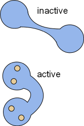 Calcium ions influxing through CaL channels and from the sarcoplasmic reticulum will binds with calmodulin, CaM
(CALcium MODULatINg). This ubiquitous protein is dumbbell-shaped when inactive but, upon activation by the binding
of four calcium ions, changes shape as shown in the illustrations to the right. In the bent form, calcium/calmodulin (Ca/CaM)
can wrap around proteins that have calmodulin binding sites. When so wrapped the activity of the target is altered.
Calcium ions influxing through CaL channels and from the sarcoplasmic reticulum will binds with calmodulin, CaM
(CALcium MODULatINg). This ubiquitous protein is dumbbell-shaped when inactive but, upon activation by the binding
of four calcium ions, changes shape as shown in the illustrations to the right. In the bent form, calcium/calmodulin (Ca/CaM)
can wrap around proteins that have calmodulin binding sites. When so wrapped the activity of the target is altered.
As shown in the above illustration, CaL channels have two such sites; one near the internal pore opening and the other further out on
the C terminus. The site nearest the pore directly binds Ca2+ and then calmodulin attaches to them and bends. The other site
binds calcium/calmodulin that has formed in the cytoplasm. These bindings cause the inactivation gate to close more rapidly
thus inhibiting the calcium current and preventing calcium overload. This closure is more rapid than than the normal voltage-dependent inactivation
described above.
Calcium-Dependent Facilitation, CDF
 A 'kinase' is an enzyme that phosphorylates its target molecule; this causes a conformational change in the target and alters
its behavior. Calcium/calmodulin-dependent kinase II (CaMKII) is a ubiquitous enzyme with a very complicated structure as seen
at the left. It is composed of twelve separate enzymes in a double stack; these enzymes are activated by the binding of Ca/CaM. The Ca/CaM
activates the kinase so that it hydrolyzes ATP retaining the phosphate group that it will add to its targets. The
above illustration shows two sites on CaL channels that CaMKII will phosphorylate (light blue circles). The
resulting phosphorylation of these channels interferes with the ability of Ca/CaM to close the inactivation gates. Thus, calcium-dependent
facilitation (CDF) extends the calcium current through CaL channels.
A 'kinase' is an enzyme that phosphorylates its target molecule; this causes a conformational change in the target and alters
its behavior. Calcium/calmodulin-dependent kinase II (CaMKII) is a ubiquitous enzyme with a very complicated structure as seen
at the left. It is composed of twelve separate enzymes in a double stack; these enzymes are activated by the binding of Ca/CaM. The Ca/CaM
activates the kinase so that it hydrolyzes ATP retaining the phosphate group that it will add to its targets. The
above illustration shows two sites on CaL channels that CaMKII will phosphorylate (light blue circles). The
resulting phosphorylation of these channels interferes with the ability of Ca/CaM to close the inactivation gates. Thus, calcium-dependent
facilitation (CDF) extends the calcium current through CaL channels.
It is noteworthy that activation of this kinase requires a high cytoplasmic concentration of calcium ions not found under
basal conditions. However, when there is a sustained, rapid heartbeat, such as during sympathetic stimulation, sufficient calcium
do accumulate in the cytoplasm. Additional, under these conditions, there is a higher than normal concentration of
phosphokinase A (PKA) that also phosphorylates CaL channels.
Updated: 2/14/2016
Continue to Repolarization
Return to previous tutorial... Phases
Return to home page