The rate at which pacemaker action potentials are produced is controlled the effects of the autonomic nervous system (ANS) on both
the membrane and calcium clocks. This involves 'first messengers' from the ANS that target membrane receptors followed by 'second messengers'
that target both membrane channels and components of the sarcoplasmic reticulum.
Autonomic Nervous System
The autonomic nervous system (ANS) increases the rate at which pacemaker action potentials are produced via its sympathetic
division (SNS)
and decreases this rate via its parasympathetic division (PSNS). It does this by releasing neurohormones from neurons that enter
the sinoatrial node (a.k.a., pacemaker) and by a hormone released into the bloodstream from the adrenal medulla.
These compounds attach to specific membrane-bound proteins called receptors. These receptors initiate a series of reactions that
modify the activity of many of the previously described ion channels and pumps.
First Messengers
Neurohormones and hormones, that bind to receptors at the outer cell surface, are called 'first messengers'.
The SNS secretes the hormone epinephrine (E) (a.k.a, adrenaline) into the bloodstream and releases the neurohormone
norepinephrine (NE) from
the endings of neurons. The neurohormone acetylcholine (ACh) is released from PSNS neuron endings. Membrane-bound
receptor proteins have binding sites for these messengers. Pacemaker cells have beta (B) receptors that bind epinephrine
and norepinephrine and muscarinic 2 (M2) receptors that bind acetylcholine.
Receptors
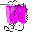 Both of the above mentioned receptors are G protein-coupled receptors (GPCRs).
These membrane-embedded proteins consist of seven alpha helices (pink) as shown in the illustration to the right. The N terminus is in the extracellular
fluid (top) and the C terminus is intracellular (bottom). Note the intracellular loop that will serve as an attachment site for
G-proteins (see below). The first messenger binds between the helices within the membrane.
Both of the above mentioned receptors are G protein-coupled receptors (GPCRs).
These membrane-embedded proteins consist of seven alpha helices (pink) as shown in the illustration to the right. The N terminus is in the extracellular
fluid (top) and the C terminus is intracellular (bottom). Note the intracellular loop that will serve as an attachment site for
G-proteins (see below). The first messenger binds between the helices within the membrane.
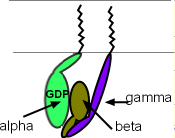 A G protein consists of three subunits as shown in the left illustration. These subunits 'dangle' into the cytoplasm
but two of them are anchored to the membrane (zig-zag lines). The alpha (green) and gamma (purple) subunits are membrane-anchored while
the beta subunit is firmly attached to the side of the gamma subunit ... forming a 'dimer' ... but is only lightly attached to the side of the alpha subunit.
Note that the alpha subunit has a molecule of guanine diphosphate
(GDP) attached ... thus the name 'G' protein. When the subunits are together as illustrated, and there is a GDP with the alpha subunit,
the G-protein can attached to the loop at the cytoplasmic
surface of the receptor as shown in the next illustration.
A G protein consists of three subunits as shown in the left illustration. These subunits 'dangle' into the cytoplasm
but two of them are anchored to the membrane (zig-zag lines). The alpha (green) and gamma (purple) subunits are membrane-anchored while
the beta subunit is firmly attached to the side of the gamma subunit ... forming a 'dimer' ... but is only lightly attached to the side of the alpha subunit.
Note that the alpha subunit has a molecule of guanine diphosphate
(GDP) attached ... thus the name 'G' protein. When the subunits are together as illustrated, and there is a GDP with the alpha subunit,
the G-protein can attached to the loop at the cytoplasmic
surface of the receptor as shown in the next illustration.
G-protein Activation
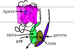 The illustration to the right shows a ligand/first messenger (blue oval) binding to the receptor. When this occurs the
GDP of the alpha subunit is replaced by a guanine triphosphate (GTP). As a result, the alpha subunit detaches from both
the receptor loop and the beta/gamma dimer . These two parts of the G protein can now diffuse away from the receptor
but they always remain just below the membrane surface because they are anchored to it. The alpha subunit's 'target' is a membrane-bound enzyme
called adenyl cyclase (AC) and the target of the beta/gamma dimer is a potassium channel; both are located nearby.
The illustration to the right shows a ligand/first messenger (blue oval) binding to the receptor. When this occurs the
GDP of the alpha subunit is replaced by a guanine triphosphate (GTP). As a result, the alpha subunit detaches from both
the receptor loop and the beta/gamma dimer . These two parts of the G protein can now diffuse away from the receptor
but they always remain just below the membrane surface because they are anchored to it. The alpha subunit's 'target' is a membrane-bound enzyme
called adenyl cyclase (AC) and the target of the beta/gamma dimer is a potassium channel; both are located nearby.
Second Messengers
The second messengers, cyclic adenosine monophosphate (cAMP) and phosphokinase A (PKA), are small molecules that are
synthesized in the cell in response to extracellular first messengers, and
diffuse through the cytoplasm to affect their targets. The subunits of G proteins are not second messengers but the alpha subunit does serve as
a 'middleman' leading to the production of second messengers.
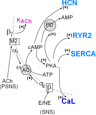 The illustration to the left, a part of the main diagram, summarizes the pathways of the autonomic nervous system involving the cardiac pacemaker.
The SNS produces first messengers epinephrine (E) and norepinephrine (NE) that bind with the membrane-bound beta
receptor .
The PSNS produces acetylcholine (ACh) that binds with the muscarinic 2 receptor (M2).
The G protein subunits (αs, αi and βγ) are shown diffusing (arrows) along the cell membrane (curved dashed line)
toward their targets. The alpha subunit targets adenyl cyclase (AC) and the beta/gamma dimer targets the KACh channel.
The illustration to the left, a part of the main diagram, summarizes the pathways of the autonomic nervous system involving the cardiac pacemaker.
The SNS produces first messengers epinephrine (E) and norepinephrine (NE) that bind with the membrane-bound beta
receptor .
The PSNS produces acetylcholine (ACh) that binds with the muscarinic 2 receptor (M2).
The G protein subunits (αs, αi and βγ) are shown diffusing (arrows) along the cell membrane (curved dashed line)
toward their targets. The alpha subunit targets adenyl cyclase (AC) and the beta/gamma dimer targets the KACh channel.
Sympathetic Division
SNS activation of adenyl cyclase causes the conversion of cytoplasmic adenosine triphosphate (ATP) to cyclic adenosine monophosphate
(cAMP). Cyclic AMP then stimulates (+) HCN channels. Cyclic AMP also activates (+) PKA (phosphokinase A) that then stimulates
(+) CaL channels, SERCA, & RYR2. Cyclic AMP is short-lived because PKA ... that it activated ... activates (+) the
enzyme phosphodiesterase (PD) and this will convert cAMP to AMP. In short, cAMP causes it's own demise.
Parasympathetic Division
When the PSNS is the more active than the SNS division, the above described second messengers (cAMP and PKA) will be in
short supply. This is because the αi will be more abundant and will decrease the activity of adenyl cyclase.
In addition, the beta/gamma subunits will open acetylcholine-activated
potassium channels (KACh) .
Updated 4/7/2015
Continue to Heart Rate Modulation
Return to previous tutorial... Calcium Clock
Return to home page