Somatic Organs
 'Somatic' means the body wall as opposed to the internal viscera. Inspection of the
diagram indicates that these organs are structures in the dermis
(arrector pili muscle of hair follicles, eccrine sweat glands and dermal arteries) and arteries
in skeletal muscles.
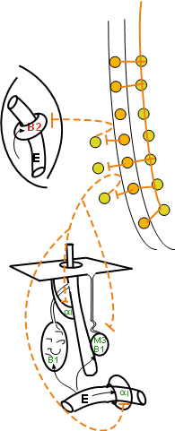
The diagram at the right shows a very short preganglionic axon (orange line) leaving
the spinal cord to synapse with a postganglionic cell body (yellow) in the ganglionic chain.
The postganglionic axon (dashed orange line) appears to curve
back to contact the spinal cord before heading to its target is in the somatic region of the body.
p>
'Somatic' means the body wall as opposed to the internal viscera. Inspection of the
diagram indicates that these organs are structures in the dermis
(arrector pili muscle of hair follicles, eccrine sweat glands and dermal arteries) and arteries
in skeletal muscles.
The diagram at the right shows a very short preganglionic axon (orange line) leaving
the spinal cord to synapse with a postganglionic cell body (yellow) in the ganglionic chain.
The postganglionic axon (dashed orange line) appears to curve
back to contact the spinal cord before heading to its target is in the somatic region of the body.
p>
The diagram at the left shows these neurons within an outline of the nerve itself. When the
preganglionic axon (orange line) leaves the cord it travels a short distance then breaks
out of the main nerve to enter a ganglion in the chain. This branch is called the white ramus.
The axon of the postganglionic neuron (dashed orange line) leaves the ganglion via another
short branch called the grey ramus. This axon re-enters the main nerve to travel to the somatic
region of the body. This seemingly unusual arrangement is characteristic of sympathetic neurons heading to the somatic region
of the body. Note the lack of parasympathetic neurons in this region.
Blood Vessels
The vast majority of somatic vessels are within skeletal muscles. The encircling smooth
muscle has beta 2 receptors -- remember that B2 receptors give a strong response to epinephrine.
Activation of beta 2 receptors causes a decrease in tone of these muscles and vasodilation that
increases blood flow. Under high sympathetic activity epinephrine diffusing from the blood
enhances this response.
Dermal Structures
Arrector Pili Muscle
These small smooth muscles originate on the lower surface of the dermis and insert on the
side of hair follicles. Follicles are not part of the actual hair but are tubular depressions
that house much of the hair. Norepinephrine activated alpha 1 receptors increases the tone of
these cells. The response is that the follicle is pulled such that the enclosed hair sticks up
straighter (hair standing on end) and some of the skin is pushed up into a bump (Goose bump).
Sweat Glands
Eccrine
The most abundant and widespread type of sweat gland is the eccrine gland. Their ducts
lead directly to a pore at the surface of the skin. These produce
a watery secretion primarily useful in thermoregulation -- evaporation from the skin surface
decrease body heat. Their innervation is quite unusual because the postganglionic neurons
secrete acetylcholine instead of the expected norepinephrine -- cholinergic sympathetic
nerves. Some texts state some vessels also have sympathetic cholinergic innervation but this
is not the case in humans. Activation of muscarinic 3 receptors on the secretory cells of
these glands causes increased sweat production.
The secretory cells of eccrine sweat glands also have beta 1 receptors. However, there is
no sympathetic adrenergic (i.e., norepinephrine) innervation and epinephrine is responsible
for their activation; sweating is increased -- 'nervous' sweat'.
Apocrine
Apocrine sweat glands are predominantly located in the armpits and groin area. Their ducts
do not lead to pores at the surface but rather into the side of hair follicles. From there
the sweat is moved to the surface. The secretion
is thick and contains many organic compounds that bacterial break down producing a foul odor.
There is no innervation to these glands. Epinephrine stimulated beta 1 receptors induce
production of this type sweat.
Last update: 10/6/2013