Inspection of the entire diagram shows "blue nerves" exiting the central nervous system from the brain and sacral spinal cord -- thus the name cranio-sacral outflow. It is also called the parasympathetic division because its activity is auxillary to the sympathetic division. The anatomy of this division is much simpler than the sympathetic division that is represented by "orange nerves" in the diagram.
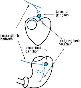
Throughout the diagram the preganglionic cell bodies (dark blue circles) give rise to axons (solid blue lines) that extend toward their targets and end as neurosecretory terminals (blue bars) that secrete acetylcholine. Most targets are postganglionic neurons; one exception is the adrenal medulla. The postganglionic cell bodies (light blue circles) lie close to, or within, the target organs. Their axons (dashed blue line) are relatively short and also end in terminals (blue bars) that secrete acetylcholine.
Ganglia are groups of postganglionic (light blue) cell bodies. This is where the preganglionic axons secrete acetylcholine onto these cell bodies. The acetylcholine binds to and activates receptors on the cell bodies. As a result, the postganglionic neurons send an impulse down their axons causing their terminals to also release acetylcholine.
Because the axons of PSNS preganglionic neurons (solid lines) are long, the location of postganglionic cell bodies are near or within the target organs. In these ganglia they synapses with postganglionic cell bodies (light blue circles) that have short axons (dashed lines) spreading throughout the organ.
When the ganglia are within a target organ they are referred to as intramural ganglia as shown in the stomach. Intramural means 'within the wall'. When the ganglia are on or near the outer surface of a target organ they are referred to as terminal as shown near the heart.
For this tutorial the term 'visceral' means all organs except those in the dermis and skeletal muscles; we'll consider those to be 'somatic'. It is not the organ itself but rather specific cell types within the organ that are innervated. These cell types are:
Last update: 10/3/2013