Throughout these tutorials the receptors that increase the activity of the cell
are color-coded green and the
those that decrease the activity of the cell are color-coded red. A more meaningful way to understand this is to
view the relationship between activation of a receptor and the response of the cell
as either direct or inverse.
A direct relationship is when a neurotransmitter binds to a receptor
and the cell's normal activity increases. For example, if the receptor is on a smooth muscle cell
it's contractile state (muscle tone) will increase. Likewise, if the neurotransmitter is not
present the cell's normal activity will decrease.
An inverse relationship is when a neurotransmitter binds to a receptor and the
cell's normal activity decreases. For example, smooth muscle tone will decrease. Likewise, if the
neurotransmitter is not present the cell's normal activity increases.
It is important to remember that both divisions of the ANS respond simultaneously although
one may override the other depending on the circumstances. It's like driving a car with
one foot on the accelerator and the other on the break at the same time!
Cholinergic Receptors
Cholinergic means "having to do with acetylcholine". The neurotransmitter acetylcholine
is released from the terminals of all preganglionic neurons in both
the sympathetic (orange) and the parasympathetic (blue) divisions of the ANS.
There are two categories of cholinergic receptors -- nicotinic and muscarinic.
Nicotinic Receptors
Acetylcholine is the neurotransmitter released from both the preganglionic and the
postganglionic neurons of the parasympathetic division.
Nicotine injected into laboratory animals causes some organs to
respond as if acetycholine had been injected. Thus, the receptors to which
both acetylcholine and nicotine can attach are called "nicotinic." There are several
subtypes of nicotinic receptors but only the N1 variety is associated with the autonomic
nervous system.
Nicotinic 1 Receptors
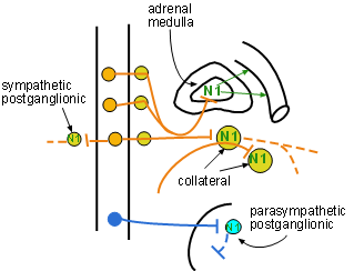
N1 receptors are located on postganglionic cell bodies in
every ANS ganglion, both sympathetic (yellow) and parasympathetic (light blue).
As implied by the green color of the N1 symbol, the response of the cell will be an
increase in its activity. These receptors are on postganglionic cell bodies and they increase
their rate of sending impulses down their axons to release their neurotransmitters. The only other
place N1 receptors are found is on chromaffin cells of the adrenal medulla . These cells
increase the amount of epinephrine and norepinephrine they secrete into the blood.
The section of the main diagram at the right shows N1 receptors on these sites. Note the
preganglionic axon (solid orange) exiting the spinal cord, passing through the ganglionic chain
without synapsing, and penetrating the adrenal cortex to reach the medulla. The large yellow circles
represent collateral ganglia. Like the connection with the adrenal medulla, preganglionic
sympathetic axons leave the cord, pass through the ganglionic chain without synapsing there and
continue on to reach postganglionic cell bodies in these ganglia. Acetylcholine released at
these sites stimulates the postganglionic neurons to release their own neurotransmitter.
On the left is the typical short sympathetic preganglionic (orange) axon. It synapses
within of the chain ganglia where it secretes acetylcholine on postganglionic cell bodies.
These cells respond by releasing their own transmitters.
Further down is a typical long parasympathetic preganglionic (dark blue) axon. It synapses
with parasympathetic postganglionic cell bodies within an intramural ganglion. These responds
by releasing their own transmitters.
Muscarinic Receptors
Muscarine, derived from the mushroom Amanita
muscaria, injected into laboratory animals causes some organs to respond
as if acetylcholine had been injected. Thus, the receptors to which
both acetylcholine and muscarine can attach are called "muscarinic."
Of the five known subtypes, M4 and M5 are restricted to the brain. The subtypes associated
with the ANS are M1, M2 and M3. Binding of acetylcholine to M1 and M3 receptors causes an increase
the activity of the cell. Binding of acetylcholine to M2 receptors causes a decrease the activity
of the cell. The main diagram shows
M1 receptors only on gastric glands lining the stomach, M2 receptors only in the heart and
M3 receptors located at numerous sites.
M1 Receptors
Acetylcholine activates M1 receptors on gastric glands increasing the secretion
of acidic 'gastric juice' into the lumen of the stomach. The relationship between acetylcholine
and the gland is direct.
M2 Receptors
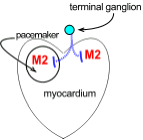 The partial diagram to the right shows M2 receptors located in two regions of the heart.
The small light blue circle near the heart represents a postganglionic parasympathetic terminal
ganglion from which axons penetrate the heart.
The circle drawn in the heart represents the cardiac
pacemaker that sets the heart rate. The rest of the heart consists of cardiac muscle,
the myocardium, that is responsible for the force of contraction.
The partial diagram to the right shows M2 receptors located in two regions of the heart.
The small light blue circle near the heart represents a postganglionic parasympathetic terminal
ganglion from which axons penetrate the heart.
The circle drawn in the heart represents the cardiac
pacemaker that sets the heart rate. The rest of the heart consists of cardiac muscle,
the myocardium, that is responsible for the force of contraction.
The relationship between acetylcholine and the response of these targets is inverse; binding of
acetylcholine to M2 receptors causes both the heart rate and the contractile force to decrease.
M3 Receptors
M3 receptors are found in many locations in the main diagram. Acetylcholine activation
of M3 receptors results in increased responses in these organs.
Responses are:
- constriction of the pupil
- increased salivation
- increased air flow
- increased gastrointestinal activity
- urinary bladder contraction
- stimulation of eccrine sweat glands.
Inspect the entire diagram to locate the organs that have M3 receptors.
Last update: 10/3/2013