Circular smooth muscle of arteries is only innervated by sympathetic fibers; the parasympathetic division is not involved. Vasoconstriction is due to of activated alpha 1 receptors. Vasodilation is due to activated beta 2 receptors.
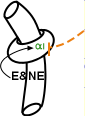
Postganglionic sympathetic neurons (orange) are scattered among the smooth muscle cells (donut) that encircle arteries. Alpha 1 receptors are characteristic of vascular smooth muscle although they are sparce in some vessels. These receptors are stimulatory -- color-coded green -- and their contraction narrows the vessel diameter thus reducing blood flow.
The adrenal hormone epinephrine E also binds to alpha 1 receptors, reaching these targets via the blood, but is less potent than norepinephrine. Notice that norepinephrine circulates in the blood in addition to being released by neurons. It enters the blood along with epinephrine from the adrenal gland.
Alpha 2 receptors -- color-coded red -- are inhibitory. Their location is unique; they are found mainly on the post-synaptic neuron terminals (orange bar) at the end of neurons innervating blood vessels of the nasal mucosa and skin. Veins have a high density of these receptors. They function to dampen the degree of vasoconstriction in these locations.
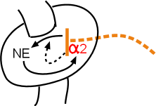
The inset to the right illustrates the details of alpha 2 receptor activation. Norepinephrine released from the terminal will bind to alpha 2 receptors (solid arrow from NE to alpha 2) on the terminal itself (orange bar). The dashed arrow pointing from the alpha 2 receptor to the arrow indicating the release of NE symbolizes the inverse relationship between alpha 2 stimulation and NE release. The overall result is a dampening of the neural signal to the vascular muscle to contract.
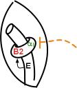
The smooth muscles of blood vessels supplying the skeletal muscles are unique because, in addition to alpha 1 receptors, they also have beta 2 receptors. These receptors are inhibitory (color-code red) indicating that, when activated, they cause relaxation of the muscle. Because these muscle fibers encircle vessels their relaxation increases vessel diameter causing increased blood flow to skeletal muscles.
Epinephrine, E is the only naturally occurring compound that activates beta 2 receptors and, even then, its potency is low. Therefore, the degree of vasodilation is dependent on the density of these receptors on the vascular muscle cells in addition to the concentration of epinephrine in the blood.
The pulmonary circuit has no autonomic innervation. It is a low-pressure circuit mainly because its arterial walls are thin. The main flow-control mechanism is due to local concentrations of dissolved respiratory gases.
The coronary circuit has no autonomic innervation. The main flow-control mechanism is due to local concentrations of metabolites and respiratory gases.
last update: 12/28/2014