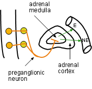
Adrenaline is the original name for what we now call epinephrine. Stimulation of the adrenal medulla by the sympathetic nervous system causes the secretion of both epinephrine (E) (80%) and norepinephrine (NE)(20%) into circulation. Epinephrine is a hormone; norepinephrine is both a hormone and a neurotransmitter.
There are two categories of receptors, alpha and beta. There are also subtypes of each. Useful generalizations concerning these are:
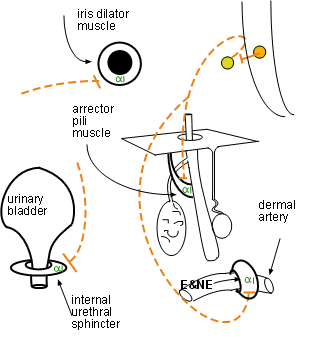
Alpha 1 receptors are more responsive to NE than to E. This is interesting as alpha 1 receptors are abundantly found on vascular smooth muscle as seen in the diagram. The relationship between the activated receptor and the cell's response is direct -- the activity (muscle tone)of the cell is increased. Alpha 1 receptors are located on:
Increase in tone of vascular smooth muscles reduces the ability of blood pressure to expand vessel diameter thus reducing blood flow to downstream organs. Increased tone in urinary and gastrointestinal sphincters reduces the passage of contents past them. Increased tone of the dilator muscle of the iris enlarges the pupil. Increased tone of arrector pili muscles pulls on the hair follicle causing hair to 'stand on end'.
Alpha 2 receptors are located on secretory terminals of some postsynaptic adrenergic neurons. When these terminals secrete norepinephrine the neurotransmitter binds with these receptors as well as with adrenergic receptors on the other side of the synapse. The presynaptic neuron's response is to decrease the amount of NE being released -- it is a negative feedback mechanism. Current research is involved with determining if alpha 2 receptors are located elsewhere.
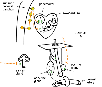
The relationship between beta 1 activation by E and/or NE is direct -- the cell's activity is increased. Beta 1 receptors are located on:
The cardiac pacemaker responds by increasing the heart rate. Simultaneously the myocardium contracts more forcefully. The physiology of the response of salivary duct cells to beta 1 activation is unclear; it appears that beta 1 activation of certain duct cells reabsorbs some water in the slowly passing saliva making it more viscous. The secretory portion of both types of sweat gland is stimulated only by E and NE from the blood, not via nerves.
Note that beta 1 activation in the heart is both neural and hormonal. In the salivary glands the activation is solely neural while in both type sweat glands it is solely due to E and NE in the blood.
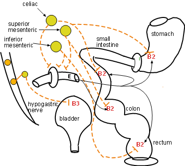
Beta 2 receptors are located on smooth muscle. The relationship between E (hormonal) / NE (neurological) activation and the response of the cell is inverse -- activity (muscle tone) of the cell is decreased. Beta 2 receptors are located on smooth muscle in:
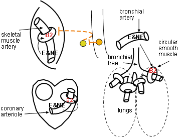
The diagram to the right emphasizes the main locations of beta 2 receptors throughout the GI tract -- stomach, small intestine, colon, rectum. Their activation results in decreased muscle tone and motility. In the urinary bladder this decreased muscle tone enables greater filling.
The diagram to the left shows relaxation of the circular smooth muscle in the bronchial tree is due to epinephrine -- note the lack of innervation at this location. Beta 2 receptors are also on small coronary arterioles thus increasing hormonally induced blood flow within the musculature of the heart. These receptors are the primary receptor in skeletal muscles arteries resulting in enhanced blood flow especially when epinephrine is present.
Inspection of the main diagram to determine which arteries have alpha 1 sites and which have beta 2 sites. Remember that epinephrine is the best activator of B2 sites.
Last update: 10/4/2013