Sympathetic Division - Thoracolumbar Outflow
Nerve Anatomy
Inspection of the entire diagram shows that these nerves contain axons forming
two-neuron chains.
The cell bodies of the first or preganglionic neurons (orange circles) are
located within the thoracolumbar region of the spinal cord in the lateral grey horns.
Their axons ( orange lines) leave the cord to synapse within ganglia with the cell bodies
of the second or postganglionic neurons (yellow circles); axons of these neurons are
shown as dashed orange lines.
Ganglia
As in the parasympathetic division the cell bodies of the postganglionic neurons are clustered
together to form ganglia. Similarly it is acetylcholine that is secreted onto these cell bodies
by the terminals of the preganglionic neurons. The postganglionic neurons respond by secreting
norepinephrine.
Organs
For this tutorial the term 'visceral' means all organs except those
in the dermis and skeletal muscles; we'll consider those to be 'somatic'.
It is not the organ itself but rather specific cell types within the organ that are innervated.
These cell types are:
smooth muscle
cardiac pacemaker cells
cardiac contractile cells
glandular cells
Visceral organs that contain these cells are shown throughout the main diagram. The type of
cell innervated within each of these organs will be described in following tutorials.
Paravertebral or Chain
Innervation of Visceral Organs
Two chains of ganglia lie along each side, i.e. paravertebral, of the spinal cord --only one is
shown in the main diagram except in the cervical (neck) region.
Each ganglion is a cluster of postganglionic cell bodies ( yellow circles).
The nerve that links the ganglia into a chain consists of hundreds of preganglionic axons
(orange lines) running up and down between the ganglia. The entire structure is called the ganglionic
chain.
The diagram at the right represents
a preganglionic axon exiting the spinal cord. It does
not synapse in the nearest ganglion but instead turns and passes through several other
ganglia before finally synapsing in one of them. A postganglionic axon (dashed
orange line) exits this ganglion to travel to a target.
Innervation of Somatic Organs
 'Somatic' means the body wall as opposed to the internal viscera. Inspection of the lower left side of the main
diagram shows the somatic region of the body where arteries within skeletal muscles and structures in the skin
(arrector pili muscle of hair follicles, eccrine sweat glands and dermal arteries) are located.
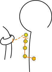
The small diagram at the left shows a preganglionic axon (orange) leaving
the spinal cord to synapse with a postganglionic cell body (yellow) in the ganglionic chain.
The postganglionic axon (dashed orange line) appears to curve
back to contact the spinal cord before heading to its target is in the somatic region of the body.
'Somatic' means the body wall as opposed to the internal viscera. Inspection of the lower left side of the main
diagram shows the somatic region of the body where arteries within skeletal muscles and structures in the skin
(arrector pili muscle of hair follicles, eccrine sweat glands and dermal arteries) are located.
The small diagram at the left shows a preganglionic axon (orange) leaving
the spinal cord to synapse with a postganglionic cell body (yellow) in the ganglionic chain.
The postganglionic axon (dashed orange line) appears to curve
back to contact the spinal cord before heading to its target is in the somatic region of the body.
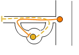 The adjacent diagram shows these neurons within an outline of the nerve itself. When the preganglionic
axon (orange line) leaves the cord it travels a short distance then breaks out of the main nerve
to enter a ganglion in the chain. This branch is called the white ramus. The axon of the
postganglionic neuron (dashed orange line) leaves the ganglion via another short branch called the
grey ramus.
This axon re-enters the main nerve to travel to the somatic region of the body.
The adjacent diagram shows these neurons within an outline of the nerve itself. When the preganglionic
axon (orange line) leaves the cord it travels a short distance then breaks out of the main nerve
to enter a ganglion in the chain. This branch is called the white ramus. The axon of the
postganglionic neuron (dashed orange line) leaves the ganglion via another short branch called the
grey ramus.
This axon re-enters the main nerve to travel to the somatic region of the body.
Innervation of the Adrenal Medulla
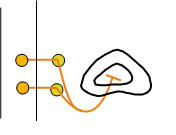 The exception to the 'two neuron rule' is the innervation of the adrenal medulla; there are no postganglionic
neurons involved...there are only preganglionic neurons (orange). Their axons
exit the cord, pass through the nearest sympathetic ganglion (yellow circle),
penetrate the cortex (i.e., outer most part) of the gland to terminate within the medulla
(i.e., inner most part) of the gland.
The exception to the 'two neuron rule' is the innervation of the adrenal medulla; there are no postganglionic
neurons involved...there are only preganglionic neurons (orange). Their axons
exit the cord, pass through the nearest sympathetic ganglion (yellow circle),
penetrate the cortex (i.e., outer most part) of the gland to terminate within the medulla
(i.e., inner most part) of the gland.
Prevertebral or Collateral
Inspection of the central area of the main diagram shows three large, yellow circles; these represent
prevertebral/collateral ganglia which are at the midline of the body near the aorta.
They are called "prevertebral" because they lie "in front of the
vertebrae." They are also called "collateral" which means "running side by side" because preganglionic neurons from both the right and
left side of the spinal cord come together in these midline ganglia.
In the main diagram, the preganglionic axons (solid orange lines)
entering these ganglia represent neurons in, from top to bottom, the greater, lesser and least
splanchnic nerves. 'Splanchnic' refers to the visceral organs; usually those below the diaphragm.
The top ganglion is called the celiac, the middle is called the
superior mesenteric, and the lower is called the inferior mesenteric.
Their names are derived from the arteries they are near.
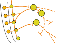
The diagram to the left depicts the basic pattern that the two-neuron pathway uses to reach targets
in the abdominal and pelvic regions. Preganglionic axons (orange line) pass
through nearby paravertebral/chain ganglia (yellow circles) after exiting the spinal cord.
Their axons are longer than typical ones and they continue toward the midline of the body
where collateral ganglia (yellow circles) are located. These preganglionic
axons are bundled together forming splanchnic nerves. Postganglionic axons
(dashed orange lines) exit the ganglia and proceed to
organs of the gastrointestinal tract and urinary bladder.
Last update: 9/27/2013